HOME

NEWS & WEATHER

UNIVERSITY

SCIENCE   General
                  My research


INDIA      General
               My pictures  
MUSIC

FAVOURITES shopping, friends,
DIY, entertainment,
Southampton etc

BOOKS

TRAVEL & HOLIDAYS

FAMILY

MEDICAL

PHOTOGRAPHY

BIRDS

SEARCH ENGINES
   All engines
   Google
   YouTube
   Stumbleupon

MY NET PROVIDER

NOTE 'My files' open with:
ppt Powerpoint
doc Word
xls Excel
pdf Acrobat Reader      [download free]
jpg Opens direct
html Opens direct;
For BIGGER text
Click Ctl plus +
Normal text click Ctl plus 0

VIDEOS can be speeded up by downloading an accelerator

HOME


BACK TO SCIENCE MAIN PAGE

         My Research and Teaching Pages

These pages contain extensive material on my own research and on my teaching

Outline CV My publications [in Word] My publications arranged by subject; with links to pdf files
Presentations used in teaching:These are PowerPoint presentations and all include links to download the original presentations if required. These were prepared for use in the University of East Anglia and Sri Venkateswara University in Tirupati, India. Topics include Introduction to Microbial metabolism, Bioenergetics, Methylotrophy
Summaries of my research
Websites I use [to data bases, literature, protein structure manipulations]
The Quinoprotein Symposium I organisised in 2002 in Southampton
Bits and pieces (for my use)


Summaries of my research
 
The Biochemistry of Methylotrophs
Growth of methylotrophs on C2 and C3 compounds
Completion of the Serine cycle in methylotrophs; a history
A History of Methanotrophy
The PQQ-containing quinoproteins
Electron transport and cytochromes
The proposed role of PQQ as a vitamin
Summary of some of my research topics as Powerpoint presentations


The Biochemistry of Methylotrophs. Microbes that are able to grow on compounds with only one carbon atom [C1-compounds] such as methane, methanol, methylamine etc are called Methylotrophs. All the earlier work on these microbes [up to 1981]is described in Anthony (1982): The Biochemistry of Methylotrophs.
My main interests are their carbon assimilation pathways and their energy transduction mechanisms.
    
The Serine Cycle has at last been completed; for the work completing the cycle click here.
    The first step in the oxidation of methanol is catalysed by the enzyme methanol dehydrogenase [MDH].
We first described this in 1964 and later showed it to be a new type of enzyme having a completely novel prosthetic group called PyrroloQuinoline Quinone [PQQ]; it was the first of a whole new family of enzymes called quinoproteins.

TOP

The PQQ-containing quinoproteins. These are the equivalent of flavoproteins that have  the riboflavin derivatives FMN or FAD as their prosthetic groups.
The quinoproteins on which we have worked are:
The Methanol Dehydrogenases [MDH] of methylotrophs;
The membrane Glucose Dehydrogenase of enteric bacteria, acetic acid bacteria and pseudomonads;
The membrane Alcohol Dehydrogenase of acetic acid bacteria.

Link to the pictorial celebration of the quinoprotein Symposium I organised in 2002 in Southampton
A major part of  my work has been working out how energy [ATP] is obtained by linking the methanol dehydrogenase in an electron transport chain to oxygen. After our discovery of PQQ [whose structure was later determined by others] it was found to be the prosthetic group of other bacterial enzymes such as glucose dehydrogenase [GDH] and we have also been involved in studies of this quinoprotein. The structure of PQQ.


     The structure and mechanism of the quinoprotein methanol dehydrogenase
Our X-ray structure of methanol dehydrogenase [MDH] was the first high resolution structure of a quinoprotein. It has a tetrameric structure. The α2β2-subunit (600 amino acids) is a superbarrel made up of eight radially-arranged β-sheets (the 'propeller fold'), held together by 'tryptophan-docking motifs. PQQ, intimately bonded to a Ca2+ ion, is buried in the interior of the superbarrel. The floor of the active site chamber is formed by a tryptophan residue and the ceiling formed by a ring structure arising from a disulphide bridge between adjacent cysteine residues joined by a novel non-planar peptide bond. This is the only example of such a structure in an active enzyme; reduction inactivates (reversibly) the enzyme but its function is unknown. It has been proposed that a catalytic base (possibly Asp303) initiates the reaction by abstraction of a proton from the alcohol substrate. The Ca2+ ion is coordinated to The C-5 carbonyl oxygen of PQQ thus facilitating polarisation of the electrophilic C-5 for subsequent attack by an oxyanion or hydride.
The remarkable 'propeller' superbarrel structure with the PQQ prosthetic group and calcium ion at its active site is best appreciated in the movie [produced with help of Stuart Findlow].


Protein Data Base [pdb] files: A2B2;  A1B1 [for pqq select pqq; for calcium ion select ca]

  The 'propeller' structure of MDH
The 'Tryptophan docking' system that holds together the propeller blades
 
TOP OF QUINOPROTEINS
TOP OF MY RESEARCH
 
     The electron transport chain from MDH to oxygen: This is very unusual as it consists of 3 periplasmic proteins and a typical cytochrome oxidase. The primary electron acceptor from MDH is a completely novel type of cytochrome called cytochrome cL; this passes electrons to a more typical small cytochrome cH which is the electron donor to the oxidase.
 
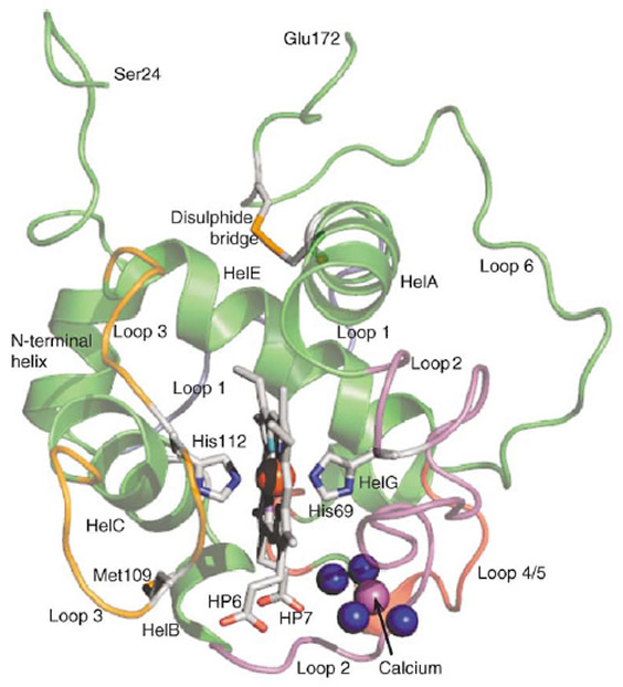
    Cytochrome cL: The structure of cytochrome cL from Methylobacterium extorquens has been determined by X-ray crystallography to a resolution of 1.6 A ° . This unusually large, acidic cytochrome is the physiological electron acceptor for the quinoprotein methanol dehydrogenase in the periplasm of methylotrophic bacteria. Its amino acid sequence is completely different from that of other cytochromes but its X-ray structure reveals a core that is typical of class I cytochromes c, having a-helices folded into a compact structure enclosing the single haem c prosthetic group and leaving one edge of the haem exposed. The haem is bound through thioether bonds to Cys65 and Cys68, and the fifth ligand to the haem iron is provided by His69. Remarkably, the sixth ligand is provided by His112, and not by Met109, which had been shown to be the sixth ligand in solution.
Cytochrome cL is unusual in having a disulphide bridge that tethers the long C-terminal extension to the body of the structure. The crystal structure reveals that, close to the inner haem propionate, there is tightly bound calcium ion that is likely to be involved in stabilization of the redox potential, and that may be important in the flow of electrons from reduced pyrroloquinoline quinone in methanol dehydrogenase to the haem of cytochrome cL. As predicted, both haem propionates are exposed to solvent, accounting for the unusual influence of pH on the redox potential of this cytochrome.

For Protein Data Bank file [pdb]: cytochrome cL
The main structural features of cytochrome cL. Although there is no sequence identity with other cytochromes, the helices A, C and E constitute the typical haem-enclosing fold seen in all cytochromes c. Helices are labelled HelA, HelB, etc. Loop 1 (grey) joins the N-terminal helix and helix A; loop 2 (purple) between helix A and helix B, carries the haem-binding sequence and the amino acid residues that coordinate to the calcium ion; loop 3 (orange) is the exceptionally flexible loop that joins helix C to helix E, and carries the sixth ligand to the haem (His112) and the methionine (Met109) that is the sixth ligand in solution. The red sphere is the iron atom at the centre of the haem prosthetic group. HP6 is the outer haem propionate group and HP7 is the inner haem propionate group. The blue spheres are the water molecules (Wat6-Wat9) that coordinate to the calcium ion (magenta sphere). Met109 is the residue that forms the sixth ligand to the haem in solutions of the cytochrome.
TOP
Cytochrome cH: This small cytochrome mediates electron flow from cytochrome cL to the oxidase. Cytochrome cH is the electron donor to the oxidase in methylotrophic bacteria. Its amino acid sequence suggests that it is a typical Class I cytochrome c, but some features of the sequence indicated that its structure might be of special interest. The structure of oxidized cytochrome cH has been solved to 2.0 Å resolution by X-ray diffraction. It has the classical tertiary structure of the Class 1 cytochromes c but bears a closer gross resemblance to mitochondrial cytochrome c than to the bacterial cytochrome c2. The left-hand side of the haem cleft is unique; in particular, it is highly hydrophobic, the usual water is absent, and the “conserved” Tyr67 is replaced by tryptophan. A number of features of the structure demonstrate that the usual hydrogen bonding network involving water in the haem channel is not essential and that other mechanisms may exist for modulation of redox potentials in this cytochrome.
For Protein Data Bank file [pdb]: Cytochrome cH

TOP

The structure and mechanism of the quinoprotein Glucose Dehydrogenase (GDH) of Escherichia coli

This quinoprotein is responsible for oxidation of glucose to gluconic acid in enteric bacteria, acetic acid bacteria and pseudomonads. It is anchored in the periplasmic membrane by the C-terminal domain made up of 5 membrane-spanning helices. the reaction is catalysed in the periplasm by the N-terminal domain; the amino acid sequence of this domain is sufficiently similar to that of methanol dehydrogenase (MDH) for its structure to be modelled using the MDH X-ray coordinates. Particularly important in achieving this were the conserved beta sheet structures that form the 'propeller blades', and the 'tryptophan-docking motifs' that hold these together [5]. Some sequence that was absent from MDH could not be modelled; this probably forms a loop [80 amino acids] that forms a domain close to the membrane which may contain the site for binding the membrane electron acceptor ubiquinone [UQ]. The active site of the GDH is very similar to that of MDH with the important difference that the novel disulphide ring in MDH is absent from GDH. This is because this ring is involved in the transfer of electrons [one at a time] from reduced PQQ in MDH to through the protein to cytochrome cL. In GDH the electrons are passed by way of a bound ubiquinone to membrane ubiquinone, which is then oxidised by a quinol oxidase.
      The model GDH has been used very successfully by ourselves and by other groups as the basis for site-directed mutagenesis of GDH [see References].

For Protein Data Base [pdb]file: GDH  H262Y mutant GDH


The 'propeller' structure of the model GDH [1] The active site of GDH [1]. The Metal may be Mg or Ca [3]
 

A

B

The active site of the model GDH.

A: The disulphide ring of MDH is replaced by His262 [1, 2]

B: His262 was replaced by Tyrosine [2]

C: View of the 'open' active site of GDH
[2]


C

 
Electron transport from membrane GDH and ADH
TOP OF QUINOPROTEINS
TOP OF MY RESEARCH

References to Glucose Dehydrogenase

1. Cozier, G.E. & Anthony, C. (1995). The structure of the quinoprotein glucose dehydrogenase of Escherichia coli modelled on that of methanol dehydrogenase from Methylobacterium extorquens. Biochemical Journal 312, 679-685.
2. Cozier, G.E., Salleh, R.A. & Anthony, C. (1999). Characterisation of the membrane glucose dehydrogenase from Escherichia coli and characterisation of a site directed mutant in which His262 has been changed to tyrosine. Biochem. J.  340, 639-647.
3. James, P.L. and Anthony, C. (2003). The metal ion in the active site of the membrane glucose dehydrogenase of Escherichia coli. Biochim. Biophys. Acta 1467, 200-205.

REVIEWS
4. Anthony, C. & Ghosh, M. (1998). The structure and function of the PQQ-containing quinoprotein dehydrogenases. Progress in Biophysics and Molecular Biology 69, 1-21.
5. Goodwin P M, Anthony C (1998) The biochemistry, physiology and genetics of
PQQ and PQQ-containing enzymes. In: Advances in Microbial Physiology.
(Poole R K eds), 40, 1-80.Academic Press, London.
6. Anthony, C. (2004) The Pyrroloquinoline Quinone (PQQ)-Containing Dehydrogenases. In Zannoni D. (ed): Respiration in Archaea and Bacteria. Vol. 1. Diversity of Prokaryotic Electron Transport Carriers, pp. 203-225. Kluwer Academic Publishers. Printed in The Netherlands.
7. Anthony, C. (2004). The quinoprotein dehydrogenases for methanol and glucose. Archives of Biochemistry and Biophysics 428, 2–9.

Publications on GDH by other important research groups
(Note: much of this work was based on our model GDH described above).

M. Yamada, K. Sumi, O. Adachi, Y. Yamada (1993), Topological analysis of
quinoprotein glucose dehydrogenase in Escherichia coli and its ubiquinone
binding site, J. Biol. Chem. 268, 12812– 12817.

M.D. Elias, M. Tanaka, H. Izu, K. Matsushita, O. Adachi, M. Yamada (2000)
Functions of amino acid residues in the active site of Escherichia
coli pyrroloquinoline quinone-containing quinoprotein glucose dehydrogenase.
J. Biol. Chem. 275, 7321– 7326.

MD. Elias, Satsuki Nakamura, Catharina T. Migita, Hideto Miyoshi, Hirohide Toyama,
Kazunobu Matsushita, Osao Adachi and Mamoru Yamada (2004). Occurrence of a Bound Ubiquinone and Its Function in Escherichia coli Membrane-bound Quinoprotein Glucose Dehydrogenase. Journal of Biological Chemistry 279, 3078 – 3083.

Mamoru Yamada, M D Elias, Kazunobu Matsushita, Catharina T Migita, Osao Adachi (2003). Escherichia coli PQQ-containing quinoprotein glucose dehydrogenase: its structure comparison with other quinoproteins. Biochimica et biophysica acta 1647, 185-92.

Kazuo Kobayashi, Golam Mustafa, Seiichi Tagawa, Mamoru Yamada (2005). Transient formation of a neutral ubisemiquinone radical and subsequent intramolecular electron transfer to pyrroloquinoline quinone in the Escherichia coli membrane-integrated glucose dehydrogenase . Biochemistry. 44, 13567-72.

Golam Mustafa, Catharina T. Migita, Yoshinori Ishikawa, Kazuo Kobayashi, Seiichi Tagawa and Mamoru Yamada (2008).  Menaquinone as Well as Ubiquinone as a Bound Quinone Crucial for Catalytic Activity and Intramolecular Electron Transfer in Escherichia coli Membrane-bound Glucose Dehydrogenase.  Journal of Biological Chemistry 283, 28169–28175.

Sode K, Yoshida H, Matsumura K, Kikuchi T, Watanabe M, Yasutake N, Ito S,
Sano H (1995) Elucidation of the region responsible for EDTA tolerance in PQQ glucose dehydrogenase by constructing Escherichia coli and Acinetobacter calcoaceticus chimeric enzymes. Biochem. Biophys. Res. Commun. 211, 268-273.

H. Yoshida, K. Sode (1996). Thr424 to Asn substitution alters bivalent
metal specificity of pyrroloquinoline quinone glucose dehydrogenase,
J. Biochem. Mol. Biol. Biophys. 1 89– 93.

H. Yoshida, K. Kojima, A.B. Witarto, K. Sode,(1999). Engineering a chimeric
pyrroloquinoline quinone glucose dehydrogenase: improvement
of EDTA tolerance, thermal stability and substrate specificity,
Protein Eng. 12 63– 70.

Goldstein A H, Lester T, Brown J (2003) Research on the metabolic engineering
of the direct oxidation pathway for extraction of phosphate from ore has generated
preliminary evidence for PQQ biosynthesis in Escherichia coli as well as a
possible role for the highly conserved region of quinoprotein dehydrogenases.
Biochem. Biophys. Acta 1647, 266-271.

Tripura C B, Podile A R, (2007) Properties of chimeric glucose dehydrogenase
improved by site directed mutagenesis. J. Biotechnol. 131, 197-204.


TOP OF QUINOPROTEINS
TOP OF MY RESEARCH
 

A model for Alcohol Dehdrogenase (ADH) of acetic acid bacteria

This model was achieved using the same methods as that used for GDH [above].
The ADH of Acetobacter aceti is a membrane quinohaemoprotein. We modelled its structure on the X-ray coordinates of methanol dehydrogenase. The basic superbarrel structure and the active-site region are retained, indicating essentially similar mechanisms of action, but there are considerable differences in the external loops, particularly those involved in formation of the shallow funnel leading to the active site.
Cozier, G.E. & Anthony, C. (1995). The structure of the quinoprotein glucose dehydrogenase of Escherichia coli modelled on that of methanol dehydrogenase from Methylobacterium extorquens. Biochemical  Journal 312, 679-685.

Protein Data Base [pdb] coordinates for ADH

'Propeller fold' structure of ADH Active site of ADH
 
Electron transport from memrane GDH and ADH

TOP OF QUINOPROTEINS
TOP OF MY RESEARCH

 
The proposed role of PQQ as a vitamin
If you came here by a link from Wikipedia you will be interested in another page on this site that discusses the Wikipedia PQQ page: go to Wikipedia and PQQ

This topic is covered also in my page on PQQ as a nutritional supplement.
The Wikipedia site on PQQ is so full of errors I have prepared a page to correct them: Wikipedia and PQQ
No mammalian PQQ-containing enzyme (quinoprotein) has been described. If such an enzyme does exist then PQQ would almost certainly be a vitamin [analogous to riboflavin, needed in the diet for production of essential flavoproteins].  Such an enzyme was reported in Nature in 2003, leading to the claim for  the discovery of the first new vitamin for 55 years. This claim was based on some basic misunderstandings of enzymes, protein structure and databases. We have been involved in refuting this claim. This was published in Nature in 2005 [Felton & Anthony].
  
   It was announced recently that PQQ is to be marketed as VitaPQQ, based on it being the 14th vitamin. This is based on false claims and so i have written the following summary of the arguments against it. This can be downloaded as a Word document.


PQQ is now to be produced as a food supplement by Mitsubishi and sold as a vitamin by Maypro

Mitsubishi Announcement: "On August 15, 2008, Mitsubishi Gas Chemical (MGC) announced that it had received official acceptance from the U.S. Food and Drug Administration (FDA) for its notification of coenzyme pyrroloquinoline quinone (PQQ) as a new dietary ingredient. Having received this acceptance, MGC will begin developing the U.S. market for PQQ as an initial step toward the commercialization of PQQ". Mitsubishi statement.

NPI Center announcement: Highly-Anticipated PQQ (Pyrroloquinoline Quinone) Finally Commercialized As VitaPQQ(TM) From Maypro Industries:
Hailed as a new vitamin by Nature1, VitaPQQ™ brand PQQ is now available from Maypro for use by vitamin and dietary supplement manufacturers.  The launch of VitaPQQ™ is a critical milestone in a decades-long effort to commercialize Pyrroloquinoline Quinone (PQQ), a newly discovered vitamin that offers significant health benefits due to its vitamin-like activity, cognitive function and antioxidant capacity.  VitaPQQ™ is manufactured utilizing a patented natural fermentation process. “
Announcement by NPI-Center.


A brief history of PQQ
PQQ as coenzyme.   
 PQQ  (Pyrroloquinoline quinone) was first discovered in the early 1960s as the novel prosthetic group (or coenzyme) of bacterial enzymes (now known as quinoproteins). Methanol dehydrogenase was one of the first of these, and was the enzyme subsequently most investigated [With Len Zatman, I discovered the enzyme in 1964 and published the purification and description of its coenzyme in 1967]. It is responsible for oxidizing methanol to formaldehyde in bacteria that grow on methane or methanol. The structure of this novel co-enzyme was determined by Olga Kennard’s group by X-ray crystallography in Cambridge University in 1979. Other quinoproteins containing PQQ include the enzyme responsible for production of vinegar from ethanol by acetic acid bacteria, and glucose dehydrogenase in (amongst others) enteric bacteria. We published the first high resolution structure of methanol dehydrogenase (using X-ray crystallography) in 1995. The unusual structure has radial symmetry and has been called a ‘propeller structure’ and this structure is shared by all the quinoproteins.
No plants or animals have PQQ-containing enzymes. If they did so then it is likely that PQQ would be a vitamin; quinoproteins having PQQ would be analogous to flavoproteins having riboflavin (vitamin B2).

PQQ as dietary supplement. PQQ is present in tiny amounts in animals and has been shown (mainly by the group of Robert Rucker) to improve growth and reproductive performance in mice fed on chemically-defined diets, but he does not claim that this shows it to be a vitamin.  However, this sort of study has led to speculation that PQQ may eventually be found to be a vitamin for humans.

False claim that PQQ is a vitamin.  Although nutritional experiments have indicated some (unknown) metabolic or nutritional role for PQQ in mammals, it cannot seriously be accepted as a vitamin until an enzyme can be shown to require it as its coenzyme. In 2003 Kasahara and Kato claim to have provided this evidence and announced ‘A new redox-cofactor vitamin for mammals' in the top science journal Nature. This was greeted with enthusiasm -  “The first new vitamin for 55 years”. However, their claim was based on sequence analysis of an enzyme, predicted to be involved in mouse lysine metabolism, which showed that part of the protein has a ‘propeller fold',  which is a feature of all PQQ-dependent dehydrogenases.  What the evidence actually suggests is that their (predicted) protein is an interesting novel protein part of which has a propeller structure; but there is no evidence that it is a PQQ-dependent dehydrogenase. In essence their argument is this: Our mouse protein has a ‘propeller’ structure [true]; All PQQ-containing proteins have a propeller structure [true]; Therefore our protein contains PQQ [false]. This is the equivalent of the well known false argument:  All trees are green; My coat is green; My coat is a tree.
       When I pointed out to the journal Nature that their high reputation was being used to justify investments of millions of dollars in the development of PQQ as a vitamin, they investigated the original paper, agreed with our objections and published our argument against it (Felton & Anthony, Nature Vol. 433, 2005). They also published (alongside ours) a paper by Rucker disagreeing with the conclusions of Kasahara and Kato on nutritional grounds, concluding “that insufficient information is available so far to state that PQQ uniquely performs an essential vitamin function in animals”.  
      It should also be noted that the enzyme which they claim to be involved in lysine breakdown does not occur in mice; it is an irreversible enzyme involved in the opposite process of lysine biosynthesis in yeast. The whole of this part of their paper is biochemical nonsense.

CONCLUSION
Mitsubishi is producing PQQ to market as a nutritional supplement, because they know that the evidence for it being a vitamin is not valid. Maypro, and journalists who like a good story, are selling it as a New  Vitamin VitaPQQ. They quote the original Nature paper but ignore the demonstration of its faultyconclusions that was subsequently  published in Nature.

References
Kasahara, T. & Kato, T. (2003).  A new redox-cofactor vitamin for mammals Nature 422, 832.
Felton, L. M. & Anthony, C. (2005). Role of PQQ as a mammalian enzyme cofactor? | VOL 433 Nature    doi:10.1038/nature03322.
Rucker, R., Storms, D., Sheets, A., Tchaparian, E. & Fascetti, A. (2005). Is pyrroloquinoline quinone a    vitamin? NATURE | VOL 433  Nature doi:10.1038/nature03323.
Anthony, C. and Zatman, L.J. (1964). The methanol-oxidizing enzyme of Pseudomonas sp. M27.    Biochemical Journal 92, 614-621.
Anthony, C. and Zatman, L.J. (1967). The microbial oxidation of methanol: The prosthetic group of alcohol    dehydrogenase of Pseudomonas sp. M27; A new oxidoreductase prosthetic group. Biochemical Journal    104, 960-969.
Ghosh, M., Anthony, C., Harlos, K., Goodwin, M.G. & Blake, C.C.F. (1995). The refined structure of the    quinoprotein methanol dehydrogenase from Methylobacterium extorquens at 1.94Å. Structure 3,    177- 187.
Anthony, C. & Ghosh, M. (1998). The structure and function of the PQQ-containing quinoprotein    dehydrogenases. Progress in Biophysics and Molecular Biology 69, 1-21.
Anthony, C. (2003). BBA Special Issue : 3rd International Symposium on Vitamin B6, PQQ, Carbonyl    catalysis and Quinoproteins (Editor). Biochim. Biophys. Acta 1647, 1-408.

Anthony, C. (2004). The quinoprotein dehydrogenases for methanol and glucose. Archives of Biochemistry    and Biophysics 428, 2–9.
For more detailed information see my lecture slides below


Slides from a Lecture on "PQQ as a vitamin? "
Given at a conference on vitamins in Osaka and to Mitsubishi scientists in Niigata

Introduction: Although nutritional experiments have indicated some (unknown) metabolic or nutritional role for PQQ in mammals, it cannot seriously be accepted as a vitamin until an enzyme can be shown to require it as its cofactor. About one year ago Kasahara and Kato claim to have provided this evidence and announced ‘A new redox-cofactor vitamin for mammals' in Nature. This was greeted with enthusiasm by Reuters news agency “The first new vitamin for 55 years”, and its exploitation by Mitsubishi seems to be underway (put vitamin and PQQ into Google for a good survey of this). However, the claim of Kasahara and Kato was based on sequence analysis of an enzyme, predicted to be involved in mouse lysine metabolism, using databases and search engines which inappropriately label beta propeller sequences as PQQ-binding sites. The ‘sites' wrongly identified by the databases do not represent PQQ-binding sites but represent the Beta -sheets that form the ‘blades' of the ‘propeller fold' which happens to be a feature of all PQQ-dependent dehydrogenases, whose main structure is a superbarrel made up of either six or eight ‘propeller blades'. What the evidence actually suggests is that their (predicted) enzyme is an interesting novel protein having an eight-bladed beta propeller structure; but there is no evidence that it is a PQQ-dependent dehydrogenase.
    There is also no evidence that this protein has any relevance to lysine metabolism.


TOP


logo design